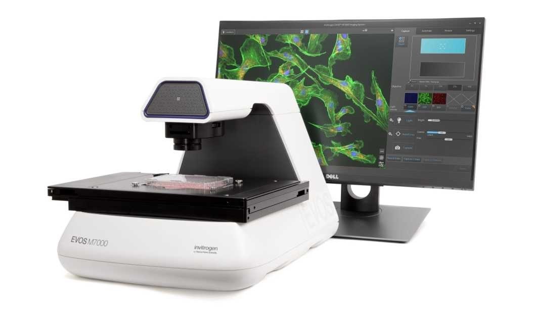


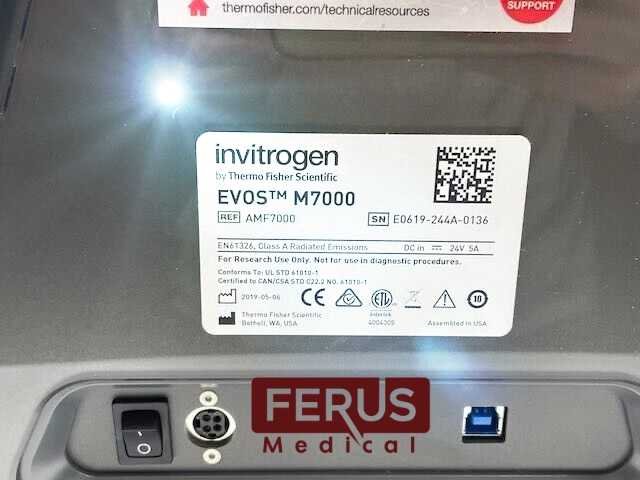


Invitrogen EVOS M7000 Imaging System
Original price was: $34,999.00.$24,999.00Current price is: $24,999.00.-29% OFF
Explore the Invitrogen EVOS M7000 Imaging System, a state-of-the-art, fully automated imaging solution designed for high-quality, multi-channel fluorescence and transmitted light imaging. Featuring high-resolution CMOS cameras, rapid data processing, and user-friendly interfaces, the EVOS M7000 is ideal for both fluorescent and colorimetric imaging in modern labs.
In stock
Description
EVOS M7000 microscope is a fully automated, personal-contact imaging system. It includes monochrome and color high-resolution CMOS cameras for the best fluorescence and colorimetric imaging. The M7000 Cell Imaging System can automatically scan multi-well plates and features fast autofocus, image acquisition, and large data processing.
The EVOS M7000 Imaging System replaces the EVOS FL Auto 2 instrument (Cat. No. AMAFD2000). It features high-resolution CMOS cameras (mono and color) with improved resolution and sensitivity across the spectrum and a high-performance PC with a responsive SSD and GPU for faster autofocusing, image acquisition and processing of large data sets.
Its user interface and software features mirror that of FL Auto 2 and it is compatible with the same accessories, including the EVOS Onstage Incubator. This system has been designed with advanced capabilities to simplify demanding cell-based imaging applications such as live-cell analysis, image tiling, and z-stacking, so you can focus on acquiring images and data rather than instrument operation.
Features of the Invitrogen EVOS M7000 Imaging System:
- High Resolution: The system offers high-resolution imaging capabilities with precision optics and advanced light cubes to visualize a wide range of fluorophores.
- Ease of Use: Like other microscopes in the EVOS line, the M7000 is designed to be user-friendly. Its intuitive interface and design allow researchers to focus on their samples, not on equipment adjustments.
- Automated Features: The system may have features such as automated stage movement, autofocus, and z-stacking capabilities to capture high-resolution 3D images.
- On-board Analysis: Some versions of the EVOS systems allow for on-board analysis, including cell counting, intensity measurements, and other image analytics.
- LED Light Cubes: These light cubes offer stable and long-lasting illumination for fluorescent imaging. They have the advantage of instant-on capability with minimal warm-up time and don’t degrade like traditional bulbs.
- Integrated Software: The EVOS M7000 might come with built-in software for image capturing, processing, and analysis.
How It Works:
- Sample Placement: Place your sample on the microscope stage.
- Selection of Parameters: Using the touchscreen interface, choose the objectives, light cubes, and other imaging parameters relevant to your sample.
- Focus and Imaging: Use the automated focus or manually adjust to get a sharp image. Once satisfied, capture the image or begin your on-board analysis.
- Image Analysis: Depending on your model and its capabilities, you can analyze the captured images directly on the system, including counting cells, measuring intensities, and more.
- Data Transfer: Most EVOS systems allow easy transfer of images and data to a computer or storage device.
The EVOS M7000 Imaging System offers these advantages:
- Outstanding image quality and versatility with a 5-position objective turret, 4-color LED fluorescence and transmitted light channels, and 3.2 MP CMOS color and B/W cameras
- Exceptional usability with fully automated X/Y scanning stage, autofocus, and acquisition routines
- High-speed image acquisition coupled with multi-position well scanning, and Z-stack and tile-stitch options lend power and flexibility to your data generation
- Fully integrated time-lapse live cell imaging using the optional EVOS Onstage Incubator for precise control of temperature, gases for normoxic or hypoxic conditions, and humidity
- Powerful image analysis capabilities for cell segmentation and quantification with the optional Celleste Image Analysis Software
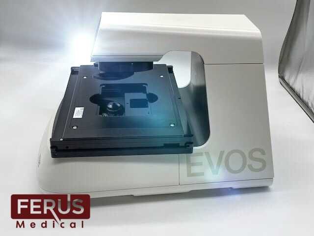
Fully automated imaging system
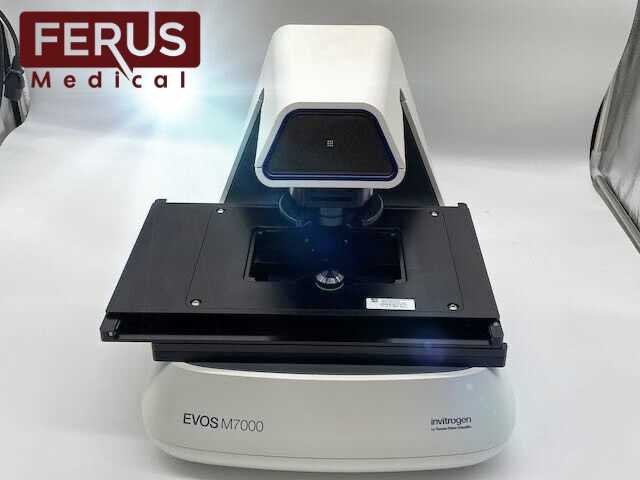
The EVOS M7000 system combines and integrates precision components with a unique modern design functionality that enables high-quality automated fluorescence imaging anywhere with unprecedented workflow efficiency. Full automation of the precision X/Y-stage movement; changing of the 4 LED fluorescent light cubes, 5-position objective turret, focus, and exposure; and switching of the dual camera make the EVOS M7000 system an exceptional platform for a variety of demanding imaging applications.
Through the intuitive acquisition interface, you can program the EVOS M7000 system to run well-plate scanning, time lapse experiments, and tile-stitch/montage area scans in Z-stack and/or time-lapse modes in your vessel of choice. For live cell imaging over multiple hours or days, the optional EVOS Onstage Incubator, which functions as an environmental chamber on top of the microscope stage, is operated seamlessly from within the instrument interface. The EVOS M7000 system is compatible with all accessories for the predecessor EVOS FL Auto imaging system.
Versatile and highly configurable
With a 5-position objective turret and the ability to simultaneous acquire images in four fluorescence channels plus transmitted light, the optical system can be easily configured to meet your needs. Choose from our broad range of high quality coverslip-corrected and long-working-distance objectives for use with plastic vessels or glass sides.
Available from 1.25X to 100X magnification in achromat, fluorite, or apochromat models, together with over twenty LED light cubes, the sample imaging options are limitless. Dual monochrome and color high-sensitivity dual CMOS cameras enable seamless imaging of both fluorescent and chromogenically stained samples. Interchangeable vessel holders accommodate most vessel types and sizes, including slides, multi-well plates, culture flasks, and Petri dishes, and afford precise control and sample alignment by the automated stage.
The vessel creation wizard in the user interface enables creation of profiles for custom sample vessels.
Powerful software
The EVOS M7000 software presents users with an extensive suite of tools for acquisition, visualization, and analysis. The user interface was designed with a strong emphasis on usability, including an option to view samples in a single field mode (Field View) or in a window that covers a much larger area (Area View).
By using a zoom function, the Area View size can be adjusted. A user can explore a large sample area at low magnification, define regions of interest, and then seamlessly transition to the Scan function in Automate Mode and execute an area scan at higher magnification.
The enhanced power of the EVOS M7000 system enables more demanding automated acquisition routines like area scanning in time lapse and Z-stacks in scan mode to be performed. Visualization capabilities include multi-field image viewing gallery, mosaic tiling and tile stitching options, Z-stack visualization, and a video recording and movie maker/editor for time lapse sequences.
The EVOS M7000 system is fully compatible with the EVOS Onstage Incubator environmental chamber for control of temperature, gases, and humidity. With the ability generate both hypoxic and normoxic conditions, it is possible to study live cells over extended periods with highly flexible time lapse automation routines.
For users needing more powerful image analysis capabilities, images acquired on the EVOS M7000 system can be analyzed directly using the optional Celleste Image Analysis Software. Celleste software offers powerful tools for image segmentation and classification that can be applied to a range of cell analysis applications for counting and measuring intensity, area, and shape changes over time, and more.
Smart LED illumination technology
All EVOS fluorescence imaging systems utilize our proprietary LED light cubes. This light engine outputs remarkable intensity over a short light path and delivers incomparable fluorophore excitation. Each light cube contains a precisely matched set of optical components to optimize the position, evenness, and intensity of the light beam. Digitally controlled LED light sources allow adjustment of illumination levels to the sample and experimental conditions to minimize the risk of photo-toxicity and photo-bleaching in time-lapse studies. Hard-coated filters give sharper edges and significantly higher transmission efficiencies than traditional soft-coated filters.
Easy to use and reliable
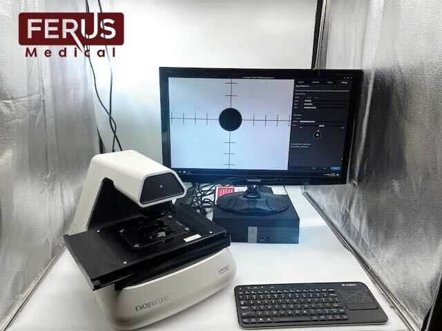
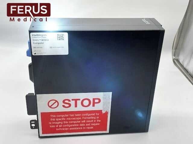
The EVOS M7000 Imaging System is powerful, yet offers plug-and-play simplicity. Like other EVOS systems, it requires no warm-up or cool-down periods. The LED light source guarantees exceptional stability and durability, so you can turn the unit on and off whenever you need to image a sample. The environmentally safe, mercury-free LED bulbs are rated for >50,000 hours, compared to 300 hours for a typical mercury bulb and 1,500 hours for a metal halide bulb. The long lifetime and low energy consumption translate into significantly lower operating costs compared to instruments with conventional light sources. An external Dell PC powers the EVOS M7000 system with an Intel Core i7-11700 processor, 32 GB DDR4 RAM, 512 GB PCIe SSD, and NVIDIA Quadro RTXA4000 with 8 GB discrete video graphics running Windows 10, designed to operate with touchscreen monitor and microscope. Whether acquiring single images or setting up automated routines, the EVOS M7000 system is remarkably easy to use and run. The 1920 x 1080 pixel resolution 23′ LCD touch screen monitor enables “finger swipes” to expand or zoom in on images, and moving between microwell plate wells is done by a single touch. Operation is also also fully controllable via mouse.
The EVOS imaging systems are built from the ground up to maximize performance and optimize workflow. You will be astonished at how easy it is to operate and amazed at how extraordinary your images look on-screen.
EVOS M7000 System and hardware specifications
- Optics:infinity‐corrected optical system; RMS‐threaded objectives with 45-mm parfocal distance
- Imaging mode:fluorescence, brightfield, color brightfield, and phase contrast
- Illumination:five position chamber for 4 fluorescence light cubes plus brightfield; light cubes with integrated hard coated filter set and LED light source with >50,000 hour life; broad selection of standard and specialty light cubes
- Imaging methods:single color, multi-color, area scan with montage or tile-stitch, time lapse, Z-stacking, movie capture
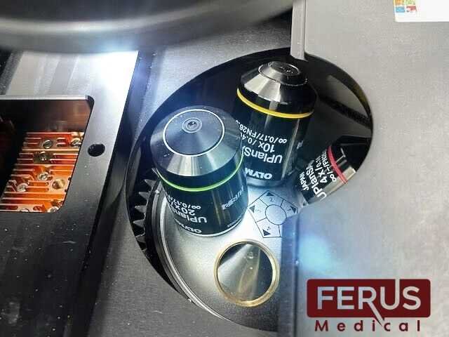
- Objective capacity:5‐position turret
- Objectives (not included):wide selection of high‐quality LWD and coverslip‐corrected objectives
- Condenser:60-mm long working distance condenser, 4 position turret with a clear aperture and 3 phase annuli
- Stage:motorized X/Y scanning stage; travel range 120 mm x 80 mm with sub‐micron resolution, drop‐in inserts to receive vessel holders and lock down holders to fix sample in place during long scans
- Focus mechanism:automated focus mechanism with sub‐micron resolution
- LCD display:23′ high‐resolution touch screen color monitor (also fully controllable via mouse); 1920 x 1080 pixel resolution
- Cameras:high-sensitivity 3.2 MP (2048 x 1536) monochrome CMOS sensor with 3.45 µm pixel resolution;high-sensitivity 3.2 MP (2048 x 1536) c
Include:
- EVOS M7000 Imaging System + Image Capture Software
- EVOS Light Cube,DAPI
- EVOS Light Cube,GFP
- EVOS 10x Objective,fluorite,LWD,
- phase-contrast,0.30NA/7.13WD
- EVOS 20x Objective,fluorite,LWD,
- phase-contrast,0.45NA/6.12WD
- EVOS 40x objective,fluorite,LWD,
- phase-contrast,0.65NA/1.79WD
- Del (Dell P2418HT) computer
- 23 inch touch screen monitor
- accessory kit
- installation/basic instruction manual
- 1 year warranty
- AC Adapter
- (4x) Filter Cubes: AMEP4650 (GFPI), AMEP4651 (GFP), AMEP4655 (TxRed), AMEP4656 (CY5)
- (3x) Plan Apochromat Objectives: AMEP4752 (4x), AMEP4753 (10x), AMEP4734 (20x)
- (3x) slide trays
- (4x) Condenser Sliders: EVOS Diffusion, Pinhole, Pinhole 4X, EVOS FL Block
- Calibration Slide
- Operating manual w/ additional USB documentation
- Shipping hardware kit
EVOS M7000 Accessory Kit
- PC w/ Evos analysis and M7000 image capture software + touchscreen monitor (Dell P2418HT)
- M7000 instrument (AMF7000) w/ AC Adapter
- (4x) Filter Cubes: AMEP4650 (GFPI), AMEP4651 (GFP), AMEP4655 (TxRed), AMEP4656 (CY5)
- (3x) Plan Apochromat Objectives: AMEP4752 (4x), AMEP4753 (10x), AMEP4734 (20x)
- (3x) slide trays
- (4x) Condenser Sliders: EVOS Diffusion, Pinhole, Pinhole 4X, EVOS FL Block
- Calibration Slide
- Operating manual w/ additional USB documentation
- Shipping hardware kit
* Note that three (3) country-specific power cord must be ordered separately in regions not using the Type B (North America) power plug:
AMEP4644 EVOS Power Cord, Type A (North America)
AMEP4645 EVOS Power Cord, Type G (United Kingdom)
AMEP4646 EVOS Power Cord, Type C (Europe)
AMEP4647 EVOS Power Cord, Type I (Australia)
AMEP4648 EVOS Power Cord, Type L (Italy)
AMEP4649 EVOS Power Cord, Type H (Israel)
AMEP4708 EVOS Power Cord, Type D (India/South Africa)
AMEP4782 Power Cord, Type I (China, CCC certified)
AMEP4786 Power Cord Type J (Switzerland)
Reviews
There are no reviews yet.