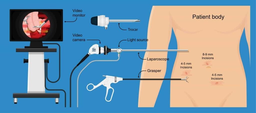
Endoscopes have been revolutionary in terms of diagnosing gastrointestinal or bronchi issues. Doctors use thin, flexible tubes with a camera at the end to look inside the body. Since its invention, endoscope video processors have revolutionized the industry, helping professionals visualize internal structures precisely.
Apart from diagnostic and therapeutic accuracy and efficacy, endoscopes have also decreased the risk of misdiagnosis, the need for exploratory surgery, and liability risks. Connecting optics, lighting, and digital image processing in real-time has made this possible.
Over the years, advanced endoscopic technology has improved. It now offers a clearer picture. This technology can connect to many tools for cauterization and sample collection. It also provides better contrast and color reproduction.
As the complexity of the medical world increases, new issues arise regularly, requiring unique diagnostic elements and clarity. At the same time, it increases the demand for minimally invasive techniques. In light of this, finding the right video endoscopy processor has become essential.
This article will explore the science and technology behind video endoscopy. We will also highlight some of the best options available to you.
Understanding Endoscope Video Processors
The idea of an endoscope is simple. It uses optical fibers, light, and image processing. The goal is to make the device as easy to move and small as possible.
Furthermore, the human body represents a uniquely extreme environment on its own. With many essential parts, pressure, fluids, and more come together. This makes it highly delicate inside.
To achieve this, a video endoscope must work well. It should show a live, transparent, and accurate image in that environment.
Video endoscopes are advanced medical tools. They have a flexible or rigid tube at the front. A high-resolution camera with a macro lens sits at the end. This macro lens can focus on objects and surfaces only 5 mm (13/64 inches) away.
The tube also features a light source to help visualize the images of internal structures for the camera. This light is usually an LED or xenon lamp.
It needs to be small but still give off enough cold light. This helps create a clear and well-lit image. You can change the cold light based on the model. This enables you to see the different things on which the camera focuses.
Video endoscopes are different from traditional fiber-optic endoscopes. Traditional endoscopes use bundles of optical fibers to send images to an eyepiece.
In contrast, video endoscopes have a digital camera at the tip. This method offers a higher resolution. The design is also lightweight and easier to process on digital units.
Endoscopy has existed since the 1800s. However, video endoscopy began in the 1960s.
Fiber-optic technology, which Basil Hirschowitz recommended, made this possible. However, it was not until the late 1980s and early 1990s that Welch Allyn combined it with CCD sensors. This allowed for direct image capture and electronic transmission.
Today, people use this concept all over the world. It follows a design pattern similar to the one from the 80s. The only improvements are in lighting, video, and profile.
How Does an Endoscope Camera Work?
The functionality of an endoscope camera hinges on the interplay between optical, mechanical, and electronic components. To elucidate, let us consider the structural elements and their roles in detail:
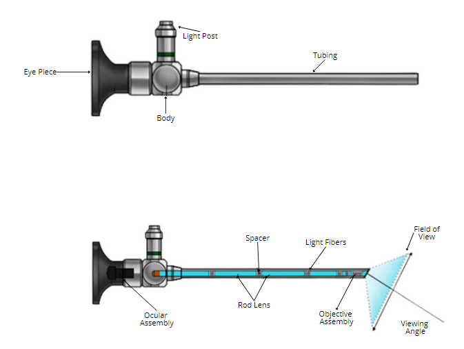
Figure 1: The Anatomy of an Endoscope
The figure above shows a schema of a commonly used endoscope. In the diagram above, the eyepiece is the front part of traditional endoscopes. Someone replaces or adds this part with an electronic interface in video endoscopes.
The light post connects to an outside light source. This is usually a xenon or LED lamp. It provides the light needed for visibility.
You can also attach other tools here. These may include biopsy forceps, suction or irrigation devices, and electrosurgical instruments.
The body houses the essential mechanical and electronic components of the endoscope. This is also where operators hold the endoscope from. The tubing at the front is the flexible or rigid pathway, through which light fibers and signal cables run. This is the part that doctors insert into the patient and extend as needed.
The field of view in the picture above shows the area you can see through the scope. The rod lens and the ocular assembly set this area. These lenses transmit light and focus the image onto the camera sensor.
The spacer keeps the optical elements aligned. The light fibers carry light from the source to the end of the endoscope.
The objective assembly has lenses at the far end. These lenses collect light from the target and focus it onto the sensor. This is the part that leads the tubing into the patient.
The angle of view, defined by the optical assembly, determines the spatial coverage of the observed region.
Moving to the inner workings of the video processing system, Figure 2 provides a schematic overview.
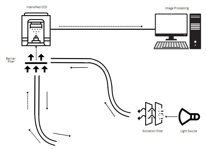
Figure 2: The Inner Workings of a Video Endoscope
Here, a 300 W xenon lamp lights up the target. The excitation filter filters the light from the lamp.
This helps isolate the right wavelength or brightness level for fluorescence endoscope systems. The filter absorbs excess light or reflects it back to the light source. Modern video endoscopes may control the lamp directly to improve energy efficiency of the system.
The light then reaches the splitting and barrier filter. This is where we direct light toward the ICCD camera.
This is also the same pathway that sends light from the camera to the image processor. The barrier here helps control the image being created. It acts as a filter for different colors and effects.
The intensified CCD camera is coupled optically with the endoscope. This allows for capturing fluorescence and visible light images with high sensitivity. Technicians apply real-time digital enhancement techniques to improve contrast, sharpness, and noise reduction, if needed. The operator may control this either using dials, buttons, or the keyboard, if any.
Finally, the system sends the signals to the personal computer, where the software analyzes the collected data. The operator then displays, stores, and assesses the processed images based on specific instructions. You can view the images in real-time or store them onto a disk as needed.
Features to Consider
Certain features are paramount to ensure optimal performance when evaluating an endoscope video processor.
- Resolution: Modern processors support resolutions up to 4K or higher, essential for discerning fine anatomical details.
- Dynamic Range: The wider the dynamic range, the more precise you can see high and low illumination regions in the same frame. This is important for complex environments or quick overviews. Some operators also adjust the filters manually to reach similar goals. However, if something was missed, the video cannot change this setting after the procedure is done. Only higher-range models do well in this area.
- Fluorescence Imaging: Higher-end video endoscopes often offer integrated fluorescence imaging capabilities. This allows for better visualization of biochemical markers such as indocyanine green (ICG).
- Auto-Enhancement Algorithms: These algorithms improve images in real-time and enhance recorded images. They enhance image quality and brightness.
- Compatibility: Different endoscopes may or may not be compatible with different video processors.
- Data integration: often involves higher-end processors directly integrating with hospital information systems (HIS) and picture archiving and communication systems (PACS). This allows for quicker record-keeping and access across the board.
How to Choose the Right Endoscope Video Processor
Selecting the right endoscope video processor requires a clear understanding of its intended use, the technical specifications needed, and the capabilities it must offer. This decision can significantly impact the efficiency of diagnostic and therapeutic procedures.
Resolution and Image Clarity
High-resolution capabilities, such as Full HD, 4K, or even 8K, can help capture minor anatomical details. The higher the resolution, the more the precision. It allows for better analysis of any anomalies. However, resolutions beyond HD may be overkill for essential diagnostic endoscopy, leading to unnecessary costs.
Dynamic Range and Contrast Management
Dynamic range defines the ability of the system, as well as diagnosticians, to differentiate between the brightest and darkest parts of an image. Advanced processors have features like TXI™ (Texture and Color Enhancement).
This helps improve surface visibility. They also include BAI-MAC™ (Brightness Adjustment Imaging) for better image contrast. This is particularly useful for identifying lesions or bleeding points across the analyzed area.
Fluorescence Imaging
Many modern processors support fluorescence imaging technologies. These include NBI™ (Narrow Band Imaging) and i-scan Optical Enhancement (OE). These technologies highlight blood vessels and mucosal structures.
They are instrumental in oncology for detecting abnormal tissues but may be redundant for clinics not performing specialized procedures.
Illumination and Light Source
LED light sources are preferred for their longevity, consistency, and lower heat production than xenon lamps. Multi-level brightness control and automatic iris adjustments are common in premium processors. This provides stable and customizable illumination, but overhead lighting control may be necessary to enhance usability in dim environments.
Ergonomics and Workflow Integration
A compact, lightweight processor like the Olympus CV-1500 simplifies setup and reduces operator fatigue. Touch-sensitive panels, pre-freeze functionality, and user-customizable modes streamline workflow. This is particularly helpful for lengthy or complex procedures.
However, it is essential to note that facilities performing high volumes of procedures benefit from such features where there is more than one operator present.
Connectivity and Data Management
Modern processors include ports for HDMI, 3G-SDI, USB, and network interfaces for real-time image sharing, storage, and remote access. Compatibility with HIS/PACS is a key feature for clinics prioritizing efficient data handling. It facilitates patient record management and team collaboration.
Clinics with limited IT infrastructure may underutilize these features and, therefore, may not require higher-end models.
Cost Efficiency and Brand Reliability
Leading manufacturers like Olympus Endoscopes, PENTAX, and KARL STORZ are well known for their product quality and features and for customer support, training, and extensive service networks.
While premium brands ensure cutting-edge features, budget-conscious facilities may find mid-range models sufficient for their needs.
Choosing the right processor depends on the intersection of clinical requirements, budget, and future-proofing. Over-investing in ultra-advanced systems may strain resources without adding value, whereas under-investing could compromise procedural outcomes.
The goal is to align capabilities with intended use, ensuring that the chosen processor enhances both diagnostic accuracy and operational efficiency.
Top 5 Video Endoscope Video Processors
Now that we’ve covered the basics of video endoscopes let’s look at the top endoscope video processors currently on the market.
1. Olympus VISERA S
The Olympus VISERA S has a built-in video diagnosis system. It provides NBI for various patient settings, like outpatient and private practices.
- Key Features: Advanced visualization, improved NBI, LED light source, pre-freeze for clear images, cybersecurity compliance.
Specifications:
- Digital Output: 3G-SDI, HDMI
- Observation Modes: WLI, NBI
- Dimensions: 308 × 157 × 461 mm
- Weight: 10.6 kg
- Power: 100–240 V AC
- Best For: Advanced diagnostics requiring enhanced visualization and minimal noise. Olympus VISERA S is suitable for urology, gynecology, and ENT use.
2. Olympus CV-1500 EVIS X1 Video System Center ($29,999.00)
The design of the EVIS X1 CV-1500 helps detect cancer early. It provides reliable and accurate diagnoses of precancerous conditions. This makes screening precise and easy.
The EVIS X1 endoscopic system uses new, easy-to-use technologies. It helps identify, evaluate, and treat diseases in the gastrointestinal tract.

- Key Features: Compact design, multiple observation modes (TXI, RDI, BAI-MAC), touch-sensitive panel, durable LED bulbs.
Specifications:
- Observation Modes: TXI, RDI, NBI, BAI-MAC
- Setup: One-touch connector, no white balance needed
- Size: Lightweight and compact
- Best For: Clinics and hospitals need versatility and streamlined workflows. Olympus CV-1500 EVIS X1 is suitable for ENT, gastroenterology, general surgery, gynecology, neurosurgery, pulmonology (bronchoscopy), thoracic surgery, urology, and more.
3. PENTAX Medical INSPIRA EPK-i8020c
The PENTAX Medical INSPIRA video processor helps improve diagnosis and treatment. It offers excellent image quality using 5 LEDs and 4K upscaling. This feature is available for PENTAX Medical endoscopes.
- Key Features: HD+/4K resolution, i-scan OE, patient data management, multiple ports for connectivity.
Specifications:
- Voltage: 100–240 V
- Dimensions: 400 × 205 × 520 mm
- Illumination: 5-LED system
- Image Enhancement: i-scan OE
- Touch screen
- Best For: High-detail imaging in advanced medical setups. PENTAX Medical INSPIRA EPK-i8020c is best suited for use in chromoendoscopy, ENT, gastroenterology, general surgery, gynecology, neurosurgery, pulmonology (bronchoscopy), thoracic surgery, urology, and more.
4. PENTAX Medical IMAGINA
The design team ingeniously tailored the PENTAX Medical IMAGINA™ video processor to facilitate day-to-day procedures.
Its compatibility with the i10c endoscope lineup ensures a smooth workflow. It offers excellent performance at an affordable price.
- Key Features: HD+, i-scan, compact design, automatic/manual brightness control.
Specifications:
- Voltage: 100–240 V
- Dimensions: 400 × 173 × 471 mm
- Image Definition: HD 1080p
- Noise Level: 39.9 dB
- Best For: Clinics prioritizing a balance of technology and cost. Suitable for use in Upper endoscopy (EGD). Colonoscopy, Endoscopic retrograde cholangiopancreatography (ERCP), Pulmonology, ENT (Ear, Nose, and Throat), and more.
5. Olympus CV-190 EVIS EXERA III ($12,999.00)
The Olympus CV-190 endoscopy system significantly advances medical imaging technology. Healthcare workers can provide the best care to their patients thanks to several features. These include high-definition imaging, NBI features, an easy-to-use design, and flexibility. Investing in the CV-190 means investing in the future of healthcare, where precision and efficiency go hand in hand.

- Key Features: Enhanced NBI, image processing, pre-freeze for clear stills, fog-free function, versatile compatibility.
Specifications:
- Output: Analog HDTV, SDTV, HD-SDI
- Dimensions: 370 × 85 × 455 mm
- Weight: 10.7 kg
- Image Enhancements: Structural and edge enhancements
- Best For: General and advanced endoscopic procedures requiring exceptional image quality. The Olympus CV-190 EVIS EXERA III best suits Gastroenterology, bronchoscopy, Urology, Gynecology, and laryngoscopy.
Here is a comparison of the top 5 endoscope video processors for endoscopic needs.
Feature/ Specification | Olympus VISERA S | Olympus CV-1500 EVIS X1 | PENTAX Medical INSPIRA EPK-i8020c | PENTAX Medical IMAGINA | Olympus CV-190 EVIS EXERA III |
Observation Modes | WLI, NBI, Contrast Enhancement | WLI, NBI, TXI, RDI, BAI-MAC | i-scan OE, HD+/4K | i-scan, HD+ | WLI, NBI |
Imaging Technology | Advanced color and depth enhancement | TXI, RDI, BAI-MAC technologies | Optical Image Enhancement (i-scan OE) | Digital Image Enhancement (i-scan SE/CE/TE) | Structural/Edge enhancement |
Light Source | LED | LED | LED | LED | LED |
Key Features | Enhanced NBI, high compatibility, clear visuals | Compact, one-touch connector, durable LEDs | Patient data management, multi-level settings | Fluoroscopy (X-Lum), noise control | Pre-freeze, fog-free function, multi-series compatibility |
Documentation & Data Management | Still images, video recording, compatibility | Touch panel, Pre-freeze, MyCV mode | USB recording, patient data management | USB recording, sub-screen freeze function | Portable memory, patient registration |
Power Supply | 100-240 V AC, 50/60 Hz, <150 VA | 100-240 V AC, 50/60 Hz, LED saves power | 100-240 V AC, 50/60 Hz | 100-240 V AC, 50/60 Hz | 100-240 V AC, 50/60 Hz, 150 VA |
Dimensions (W x H x D) | 308 x 157 x 461 mm | Compact and lightweight | 400 x 205 x 520 mm | 400 x 173 x 471 mm | 370 x 85 x 455 mm |
Weight | 10.6 kg | Lightweight | – | – | 10.7 kg |
Compatibility | CYF-VH, CH-S200-08-LB, URF-V3 | 1100 series endoscopes | Wide port variety, multi-use setup | CMOS chip system | EVIS series scopes, multi-generation use |
Image Recording Format | TIFF, JPEG, MOV | TIFF, JPEG, MOV | USB flash memory | USB flash memory | TIFF, JPEG |
Advanced Safety Features | FDA, IEC81001-5-1 compliance | LED bulbs reduce downtime | ISO 7779 acoustic noise control | Forced air cooling | Waterproof, reset-to-default settings |
Zoom Options | None | None | Yes (up to x2.0) | Yes (up to x2.0) | Yes |
Conclusion: Precision in Endoscopy with Advanced Video Processors
Endoscope video processors have become reliable and integral to modern diagnostics and therapy. Since the 1980s, they have evolved but cannot depict complex and hard-to-reach areas. They also struggle to provide different viewing angles and filters. As a result, they provide unmatched precision in high definition, leading to better diagnoses.
This list includes endoscope video processors that suit different needs.
- The Olympus VISERA S excels in advanced diagnostic settings with its noise reduction and visualization features.
- The Olympus CV-1500 is perfect for clinics needing versatile modes and efficient workflows.
- The PENTAX INSPIRA EPK-i8020c shines in high-detail imaging, making it ideal for complex cases.
- The PENTAX IMAGINA offers a cost-effective solution with excellent HD capabilities.
- The Olympus CV-190 is a well-rounded option for both general and specialized procedures.
Choosing the best one depends on your specific requirements, such as image quality, ease of use, or budget.
If you’re unsure which endoscope video processor best meets your needs, Ferus Medical is here to help. Check out our store to find exactly what you‘re looking for, or call us directly to discuss your options!
One Reply to “Top 5 Endoscope Video Processors for Endoscopic Needs”
Video Endoscopy Equipment: A Doctor's Guide for Medical Practices
[…] Endoscope Video Processors is a long, thin tube that is inserted into the body via the esophagus or anus to detect the internal organs of the body. Endoscopy is usually done in conditions like gastroesophageal reflux disease to check cancer of the esophagus or perform colonoscopies. […]